Peripheral arterial disease in non-diabetic patients tends to concentrate in the larger arteries above the knee, producing classic calf pain on walking. In diabetic patients, the disease pattern is often different — blockages cluster in the smaller tibial and peroneal arteries below the knee, frequently in more than one vessel at once, and can progress with far fewer warning symptoms. This distinction is central to how a diabetic foot surgeon in Delhi decides whether a wound will heal on its own or whether the blood supply itself needs to be restored first.
Why Diabetes Shifts Disease Below the Knee
Long-standing diabetes accelerates atherosclerosis and also causes medial arterial calcification, a stiffening of the vessel wall that is distinct from typical plaque build-up. Both processes tend to affect the tibial and peroneal arteries running from the knee to the foot more heavily than the larger vessels above it, and disease is frequently present in more than one of these vessels at the same time. Collateral blood vessels, which normally compensate for a single blockage, are less able to compensate when several vessels are affected together, leaving the foot with a thin margin of blood supply even before an ulcer or injury occurs.
How Below-Knee Disease Threatens the Foot
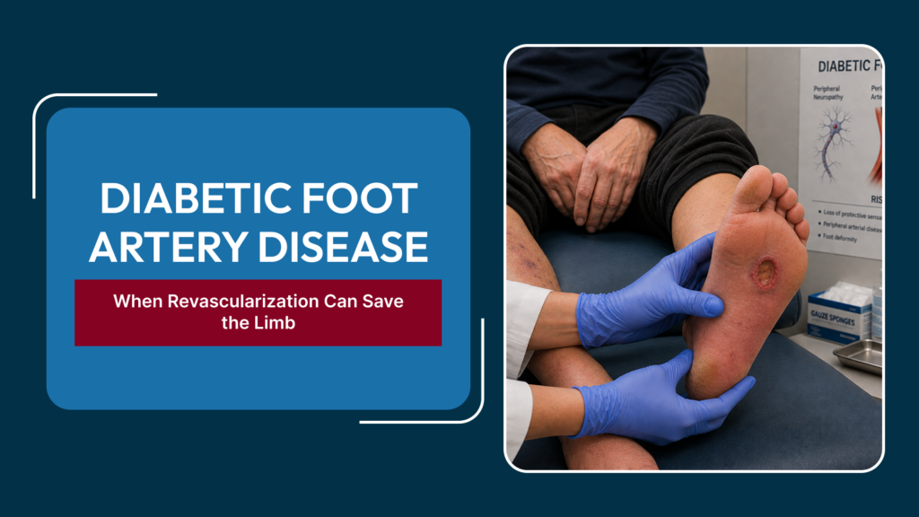
When blood flow to the foot falls below what tissue needs to survive, the result is chronic limb-threatening ischemia (CLTI) — a state marked by rest pain, non-healing ulcers, or gangrene. Because diabetic neuropathy often blunts pain sensation, patients may not feel the warning signs that would normally prompt an earlier visit, so a minor cut or callus can progress to a non-healing wound before it is even noticed. Without adequate blood flow, even meticulous wound care and infection control cannot make an ulcer heal — the tissue simply does not have the oxygen and nutrients it needs to repair itself.
Diagnosing Below-Knee Disease
The standard ankle-brachial index (ABI) can be falsely reassuring in diabetic patients because calcified vessels resist compression and give artificially high readings. A toe-brachial index, which measures pressure in the smaller toe arteries that calcify less, is often more reliable. Duplex ultrasound and CT or catheter-based angiography are then used to map exactly which tibial and peroneal vessels are blocked and how far the disease extends toward the foot, information that directly shapes the revascularization plan.
When Revascularization Becomes Necessary
Not every diabetic patient with reduced blood flow needs an intervention immediately — many are managed with risk-factor control and monitoring. Revascularization becomes necessary once a patient develops rest pain, a non-healing ulcer, or gangrene, the hallmark features of CLTI. Clinical scoring systems that weigh the size of the wound, the degree of infection, and the severity of ischemia (commonly the WIfI classification: Wound, Ischemia, foot Infection) help decide how urgently blood flow needs to be restored and whether the limb is realistically salvageable.
Revascularization Options
Endovascular Angioplasty
Balloon angioplasty, sometimes with a drug-coated balloon, is threaded through the blocked tibial artery to reopen it from within. It is well suited to shorter blockages and to patients who may not tolerate open surgery, and it can often be performed with a short recovery.
Distal Bypass Surgery
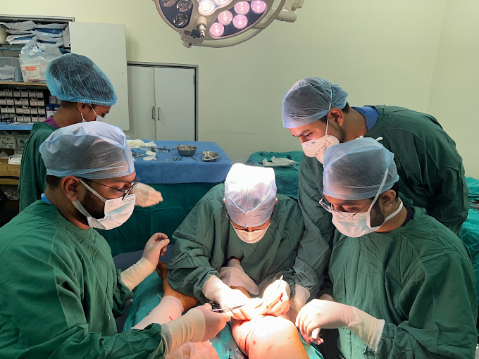
For long occlusions or heavily calcified vessels that angioplasty cannot reliably treat, a bypass using the patient’s own vein routes blood around the blockage directly to a tibial or even a pedal (foot-level) artery. Distal bypass demands a suitable vein and a patient fit for surgery, but it can achieve durable, high-volume flow to the foot when endovascular options are limited.
Angiosome-Directed Treatment
The foot is supplied by distinct vascular territories, or angiosomes, each fed by a specific artery. Where possible, revascularization is directed at the artery that most directly supplies the area of the wound, since restoring flow to the correct angiosome improves the chance of healing compared with simply reopening whichever vessel is easiest to reach.
After Revascularization
Restoring blood flow is only the first step — the wound still needs debridement of dead tissue, infection control, offloading of pressure, and regular dressing changes to heal. Blood flow is monitored after the procedure to confirm the artery has stayed open, and blood sugar and cardiovascular risk factors are managed alongside wound care, since below-knee arterial disease is a marker of more widespread vascular disease that needs ongoing attention.
Why Multi-Vessel Disease Complicates Planning
It is common for a diabetic patient to have significant disease in two or all three of the below-knee arteries at once, rather than a single isolated blockage. Planning a revascularization in this setting means deciding not just whether to treat, but which vessel offers the best combination of technical success and the most direct supply to the wound — treating the ‘easiest’ vessel is not always the one that helps the wound heal fastest. This is why detailed angiographic mapping down to the level of the foot, rather than a general assessment of ‘reduced flow,’ is central to planning below-knee revascularization.
The Cost of Delay
Because diabetic neuropathy can mask pain, patients sometimes present later than they would with a painful condition, by which point infection has often set in alongside the poor blood supply. Delay allows a limited, treatable wound to progress to deeper tissue loss or bone infection, narrowing the options from limb-salvage revascularization to a higher-level amputation. Regular foot checks and prompt attention to any new wound, however small it looks, remain the most effective way to keep below-knee arterial disease from reaching that point.
Frequently Asked Questions
Why do diabetic patients get blockages below the knee more than elsewhere?
Diabetes accelerates atherosclerosis and causes stiffening of the smaller tibial and peroneal arteries, and these vessels are also less able to develop collateral flow when more than one is affected, which together shift the disease burden below the knee.
Can a diabetic foot ulcer heal without revascularization?
If blood flow is severely reduced, wound care alone is usually not enough — the tissue needs adequate blood supply to repair itself, so restoring flow through angioplasty or bypass is often a prerequisite for healing.
Is angioplasty or bypass surgery preferred for below-knee disease?
The choice depends on the length and pattern of the blockage, the quality of the patient’s own vein, and overall fitness for surgery — shorter lesions often favor angioplasty, while long occlusions with a good vein available often favor bypass.
How is the correct artery for revascularization chosen?
Angiography maps which arteries are blocked, and where possible the artery directly supplying the wound’s angiosome, or vascular territory, is targeted to give the wound the best chance of healing.
How often should diabetic patients have their foot circulation checked?
Diabetic patients, especially those with neuropathy, benefit from at least an annual foot and pulse examination, with earlier evaluation for any new wound, discoloration, or pain that does not resolve quickly.
A non-healing diabetic foot wound is a sign the blood supply needs evaluation, not just dressing changes. Explore limb-saving revascularization options at Best Diabetic Foot Surgeon in Delhi