Every week, at least two or three patients walk into the hospital who have been living with what they assumed were “just varicose veins” — and turn out to have something considerably more serious. The reverse also happens: patients who panic about DVT turn out to have superficial venous disease that can be treated as an outpatient. Both mistakes have real consequences. This article is about learning the difference.
Your legs are telling you something. Swelling, aching, heaviness, skin discolouration, visible veins, pain that worsens as the day goes on — these are all legitimate warning signals from your venous system. But they are not all the same warning, and they do not all call for the same response. As a Vascular Surgeon In Delhi who evaluates both varicose veins and DVT as primary conditions, I see the confusion these symptoms cause — and the harm that results when patients either dismiss them or catastrophise them without proper diagnosis.
This article will walk you through what each condition actually looks like from a clinical perspective, why they are frequently confused, what distinguishes them anatomically, how we investigate and treat them differently, and when — critically — they can exist together and complicate each other.
The Anatomy Behind the Confusion
The leg has two venous systems, and understanding this is the key to understanding why these conditions get mixed up. The superficial venous system sits just under the skin — these are the veins you can see and feel. The deep venous system runs through the muscles, deep inside the leg, and is invisible from the outside. Varicose veins are a disease of the superficial system. DVT is a disease of the deep system. They share the same anatomical neighbourhood, some of the same symptoms, and critically, the same territory — which is why distinguishing them requires imaging, not just looking at the leg.
The two systems are connected by perforator veins — short vessels that run through the muscle fascia connecting deep to superficial. When the deep system is blocked by a DVT clot, the blood tries to reroute through the perforators and superficial veins. This means a new DVT can actually cause superficial varicosities to appear or worsen rapidly — a pattern that experienced specialists recognise as a red flag requiring urgent investigation.
What Varicose Veins Actually Are — and What They Are Not
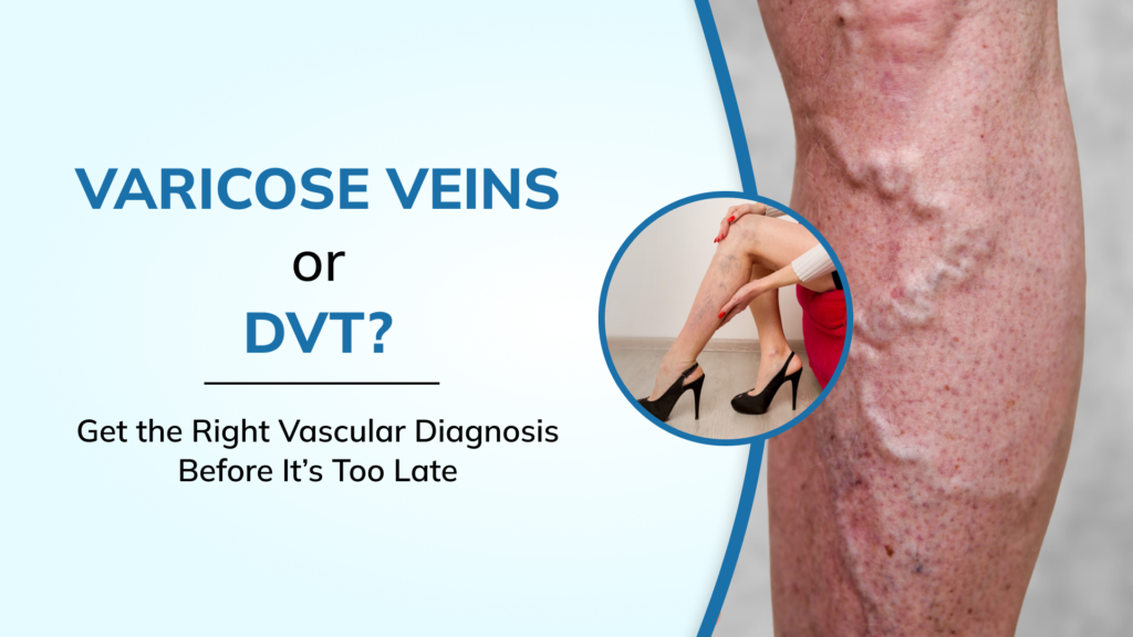
Varicose veins are dilated, twisted, elongated superficial veins caused by valve failure. Inside every vein, a series of one-way valves prevents blood from refluxing backward (downward, in the leg) between heartbeats. When these valves fail — due to genetics, prolonged standing, pregnancy, obesity, or age — blood pools in the superficial veins, which gradually stretch and dilate under the increased pressure. The result is the rope-like, bulging veins visible under the skin, usually in the back of the calf or the inner thigh.
The symptoms of varicose veins are typically worse at the end of the day and improve overnight with leg elevation. This pattern reflects gravity: blood accumulates in the legs over the course of the day when the valves cannot move it effectively upward. Classic symptoms include visible bulging veins, a dull aching or heaviness in the legs, itching over the vein, mild swelling at the ankle by evening, and in advanced cases, skin changes — brown discolouration, thickening, and in the most severe cases, venous ulcers that refuse to heal.
Varicose veins are not a cosmetic problem in their advanced form. They represent chronic venous insufficiency — a progressive condition that, if left untreated over years, can lead to severe skin damage and limb-threatening ulceration. Consulting a skilled Varicose Veins Doctor In Delhi early dramatically reduces the risk of reaching that endpoint. Treatment is highly effective and largely minimally invasive — options include endovenous laser therapy (EVLT), radiofrequency ablation (RFA), VenaSeal glue closure, foam sclerotherapy, and in certain anatomically complex cases, a combination approach. All are outpatient procedures. Most patients return to normal activity within a day or two.
“A patient comes to me with varicose veins they have had for seven years. They waited because they thought it was cosmetic. By the time they arrive, they have a venous ulcer on the inner ankle that has been there for four months, treated by three different dermatologists as a skin infection. The vein was the source the entire time. Varicose veins are a medical condition, and they progress.”
What DVT Looks Like — and Why It Feels Similar but Is Fundamentally Different
Deep vein thrombosis begins in the deep venous system, which carries roughly 90% of the blood draining from the leg. A clot in this system does not produce visible veins. What it produces is a combination of: sudden onset swelling — often in the entire leg below the knee, or the entire leg if the clot is in the thigh or pelvis — warmth, redness or cyanosis of the skin, and a deep, aching or cramping pain that is distinctly different from the tired heaviness of varicose veins. DVT pain often feels like a persistent, non-positional ache — it is there when you sit down, when you lie down, when you put the leg up. It does not resolve overnight the way varicose vein discomfort does.
The swelling in DVT is characteristically pitting — pressing firmly on the swollen area leaves a dimple that slowly fills back in. The swelling in varicose vein disease is typically non-pitting and limited to the ankle area rather than the whole leg. However — and this is important — these are tendencies, not rules. An experienced Best Vascular Surgeon In Delhi will not diagnose or exclude DVT on clinical grounds alone. The only reliable way to know is a duplex ultrasound of the leg veins, performed by a trained vascular sonographer.
The Three Scenarios Where These Conditions Genuinely Overlap
Scenario 1: Superficial Thrombophlebitis Masquerading as Varicose Veins
When a varicose vein becomes inflamed and clotted — a condition called superficial thrombophlebitis — patients feel a tender, red, hot cord along the path of the vein. This is extremely painful and alarming in appearance, but the clot is in the superficial system and rarely travels to the lungs. However, approximately 25% of cases of superficial thrombophlebitis extend to the junction where the superficial system meets the deep system — the sapheno-femoral junction — and at that point, DVT risk becomes real. A duplex ultrasound is needed to map the clot and determine proximity to the deep system. This is a scenario where what looks like an inflamed varicose vein can, in a minority of cases, be the entry point for a DVT. Proper imaging by a DVT Specialist in Delhi is non-negotiable in these cases.
Scenario 2: DVT Presenting with Visible Veins
A fresh, extensive DVT blocking the deep system forces the venous blood to reroute through collateral pathways — including the superficial system. In these cases, patients may notice new, prominent superficial veins appearing rapidly across the thigh or groin area. These are not varicose veins in the classical sense — they are collateral veins dilating under the pressure of a blocked deep system. The appearance can genuinely mimic varicose vein disease. The key distinguishing feature here is the time course: varicose veins develop over years. A new pattern of visible superficial veins appearing over days or weeks, combined with leg swelling, is a DVT until proven otherwise. This warrants same-day investigation, not a routine outpatient appointment three weeks later.
Scenario 3: Post-DVT Syndrome Causing Varicose Vein-Like Symptoms
When a DVT heals incompletely — as it often does when treatment is delayed or suboptimal — the damaged deep vein valves fail, causing venous reflux and high pressures in the deep system. This pressure is transmitted through the perforators to the superficial system, which then dilates. The result is a clinical picture almost identical to varicose vein disease: leg aching, swelling, skin discolouration, and visible superficial veins. But the underlying cause is a scarred deep venous system, not primary valve failure in the superficial veins. Treating this as standard varicose veins — ablating the superficial veins — without first addressing the deep system can worsen the situation significantly, because those superficial veins may be the only functional drainage pathway left. This is why the history of a previous DVT, however distant, changes everything about how varicose vein disease is investigated and managed.
A Best Vascular Surgeon In New Delhi will always take a thorough DVT history before planning varicose vein treatment, and will request a comprehensive duplex assessment of both the deep and superficial systems — not just a quick scan of the visible veins.
How We Investigate Leg Vein Symptoms in 2026
The gold standard first-line investigation for any symptomatic leg vein condition is a colour duplex ultrasound — a combination of B-mode imaging (which shows the structure of the vein) and Doppler flow analysis (which shows the direction and pattern of blood flow). In experienced hands, this single test can diagnose DVT with 95%+ sensitivity in the thigh veins, map varicose vein reflux patterns to plan treatment, identify perforator incompetence, and assess the valve function of the deep venous system.
CT venography or MRI venography is added when the pelvic or abdominal veins need assessment — areas that duplex cannot adequately image. These are particularly important when a patient has iliofemoral DVT (clot extending into the iliac veins or inferior vena cava), where the treatment approach — and the need for mechanical clot suction or venous stenting — depends on the full anatomical picture.
Blood tests including D-dimer, full blood count, renal function, and in selected patients a full thrombophilia screen are ordered based on clinical context. The entire diagnostic workup, from first consultation to confirmed diagnosis, typically takes one clinical visit for straightforward cases. Complex cases — those with bilateral involvement, pelvic symptoms, or atypical presentations — may require multimodality imaging over 24 to 48 hours. You can find detailed answers to questions about investigation, wait times, and what to bring to your first appointment at the vascular surgery FAQs page.
Treatment: Why These Two Conditions Require Completely Different Specialists
Varicose veins are managed with thermal or non-thermal ablation of the refluxing superficial trunk veins. The great saphenous vein running up the inner leg and the small saphenous vein running up the back of the calf are the two most common targets. EVLT (endovenous laser therapy) uses laser energy delivered through a thin fibre placed inside the vein to close it from the inside out. Radiofrequency ablation uses heat generated by radio waves. VenaSeal uses a medical-grade cyanoacrylate glue to permanently seal the vein without heat — making it ideal for patients on anticoagulation or those in whom thermal techniques carry risk. Foam sclerotherapy injects a chemical irritant as a foam, which destroys the vein wall and scleroses it closed. All of these are outpatient procedures taking 30 to 60 minutes, requiring no general anaesthesia.
DVT management, as discussed in our detailed guide on DVT Treatment In Delhi, spans anticoagulation at one end of the spectrum to catheter-directed thrombolysis and mechanical thrombectomy — using systems like the Angiojet, Penumbra CAT 16, and Inari FlowTriever — at the other. The selection between these options depends on clot location, age, patient factors, and the treating physician’s technical capability.
What these two treatment pathways share is that they both require a specialist who understands the complete venous system — not just one part of it. An Endovascular Surgeon In Delhi trained in both superficial and deep venous disease is best positioned to give you a complete picture and a treatment plan that addresses the actual source of your problem, not just its most visible manifestation.
The Question That Tells You Everything: When Did Your Symptoms Start?
In my clinic, the single most informative question I ask a patient with leg symptoms is: did this come on over years, or over days? Varicose vein disease develops slowly, over months and years of gradual valve deterioration. The patient typically says: “I’ve had these veins for as long as I can remember — they’ve just gotten worse.” DVT, by contrast, has a beginning that the patient can usually identify: “My leg was normal last week. Three days ago it started swelling and now I can’t fit my usual shoes on.”
This time course is the first and most important filter. Combine it with the pattern of swelling (ankle-only versus whole-leg), the presence or absence of visible veins (which tells us superficial versus deep), and the response to elevation (varicose symptoms improve with overnight rest; DVT swelling does not), and you have a working clinical hypothesis before any imaging is done. Imaging then confirms or refines that hypothesis — it does not replace clinical reasoning.
When to Seek Immediate Care Versus a Scheduled Appointment
Seek same-day emergency evaluation — do not wait for a regular appointment — if you have: sudden whole-leg swelling that appeared within 24 to 48 hours; leg symptoms combined with any difficulty breathing, chest pain, or rapid heart rate; a blue or purple discolouration of the entire leg; a history of cancer, recent surgery within the last three months, or prolonged immobilisation, combined with new leg swelling. These presentations have a high probability of DVT or pulmonary embolism and need to be treated as emergencies.
A scheduled consultation with a Vascular Surgeon In New Delhi is appropriate for: long-standing varicose veins causing discomfort but no sudden worsening; spider veins or reticular veins you want assessed; leg heaviness and swelling that follows a clear daily pattern, worsens in heat and improves overnight; or a family history of varicose veins or DVT that you want evaluated proactively. For information on the breadth of vascular conditions and treatment approaches available, including aortic aneurysm management and arteriovenous malformation care, the vascular surgeon category on our website provides a comprehensive overview of the specialty.
You can also explore condition-specific resources for varicose veins and review before-and-after outcomes in the surgery gallery to understand what treatment results actually look like in real patients.
The Bottom Line: Do Not Self-Diagnose Leg Vein Problems
The overlap between varicose veins and DVT is real, clinically meaningful, and consequential. Treating a DVT like varicose veins — ignoring it, assuming it will resolve, avoiding medical care — is a decision that can end in a pulmonary embolism. Treating varicose veins like a DVT — taking anticoagulants unnecessarily, avoiding normal activity — carries its own risks of bleeding and recurrence. Both mistakes happen when patients try to self-diagnose from symptom descriptions online, or when generalist physicians manage vascular conditions without subspecialty training or access to proper imaging.
The only safe path is a duplex ultrasound, read by a trained vascular specialist who understands the full spectrum of venous disease. If you are in Delhi and dealing with any of the symptoms described in this article — swollen legs, painful visible veins, sudden leg changes, or a family history of vascular disease — the contact page is where to start. You can learn more about the full scope of care and surgical expertise at bestvascularsurgeondelhi.com — and make the decision that your leg symptoms have been waiting for you to make.