
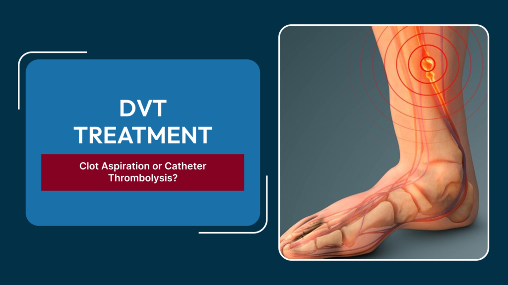
Deep vein thrombosis (DVT) is usually treated with blood thinners alone, but a subset of patients with extensive, symptomatic clots benefit from active clot removal rather than anticoagulation on its own. Two catheter-based techniques dominate this space: catheter-directed thrombolysis (CDT) and percutaneous clot aspiration (mechanical thrombectomy). Both are performed through a small puncture rather than open surgery, yet they work on different principles, carry different risks, and suit different patients. Understanding how a DVT specialist in Delhi chooses between them helps patients ask the right questions before consenting to a procedure.
Why Clot Removal Is Considered at All
Anticoagulation prevents a clot from growing and reduces the risk of pulmonary embolism, but it does not physically remove existing thrombus. Left alone, an extensive iliofemoral DVT — one that blocks the vein from the groin up into the pelvis — can lead to chronic venous hypertension, permanent valve damage, and post-thrombotic syndrome, a condition marked by lifelong leg swelling, skin changes, and in some cases ulceration. Active clot removal is generally reserved for patients with recent-onset, extensive proximal DVT, significant limb swelling or pain, a low bleeding risk, and reasonable life expectancy and mobility to benefit from the procedure.
Catheter-Directed Thrombolysis (CDT)
In CDT, a thin catheter with multiple side holes is threaded into the thrombosed segment under imaging guidance. A clot-dissolving drug — typically a tissue plasminogen activator — is then infused directly into the clot over 12 to 24 hours, sometimes longer. The patient stays in a monitored bed during the infusion, and repeat venograms check how much clot has dissolved so the dose and duration can be adjusted.
Because the drug is delivered at the site of the clot rather than systemically, CDT achieves higher local concentrations with a lower bleeding risk than whole-body thrombolysis, though bleeding — at the puncture site, or less commonly at distant sites such as the brain or gastrointestinal tract — remains the main safety concern. CDT is well suited to fresh, extensive clot burden where the goal is thorough vein clearance and preservation of valve function.
Percutaneous Clot Aspiration and Mechanical Thrombectomy
Clot aspiration uses a catheter-based device to physically suction or macerate the thrombus and remove it, often in a single session without an overnight drug infusion. Some systems combine aspiration with mechanical fragmentation; newer devices achieve large-volume clot removal with minimal or no thrombolytic drug at all, which shortens the procedure and can avoid an ICU stay.
The appeal of aspiration is speed and a lower bleeding profile, since little or no lytic drug is used. It can also be used in patients who have a relative contraindication to thrombolytic drugs, such as recent surgery or a history of bleeding. The trade-off is that aspiration alone may leave more residual clot behind in very long or organized (older) thrombus compared with a thorough thrombolytic infusion, sometimes requiring the two techniques to be combined.
How the Decision Is Made
Age and Consistency of the Clot
Fresh clot (typically under 14 days old) responds well to both techniques. Older, organized clot is firmer and often resists aspiration, making thrombolysis or a combined approach more effective.
Bleeding Risk
Patients with recent surgery, active bleeding, uncontrolled hypertension, or a history of intracranial bleeding are steered toward aspiration-based or drug-minimizing techniques rather than a prolonged thrombolytic infusion.
Extent and Location of Disease
Isolated iliofemoral disease responds well to either approach; disease extending into smaller calf veins is harder to clear with aspiration devices sized for larger vessels, and may be left to anticoagulation alone.
Underlying Anatomic Lesions
Left-sided iliofemoral DVT is frequently associated with May-Thurner syndrome, where the left iliac vein is compressed by the overlying artery. When this is found on imaging, clot removal is typically followed by venous stenting to correct the underlying compression and reduce the chance of recurrence.
Patient Factors
Symptom duration, kidney function (relevant to contrast dye load), overall mobility, and how much the patient stands to gain in quality of life all factor into the final recommendation, which is made jointly with the patient after reviewing imaging.
What Happens After the Procedure
Regardless of which technique is used, patients are typically kept on anticoagulation for several months afterward to prevent re-clotting, fitted with graduated compression stockings to support venous return, and encouraged to walk early rather than remain immobile. Follow-up duplex ultrasound checks vein patency and valve function over the following weeks and months, and any residual narrowing found on imaging may be addressed with balloon angioplasty or a stent.
Risks Common to Both Techniques
Because both CDT and aspiration thrombectomy involve catheter access to a major vein, shared risks include bleeding or bruising at the puncture site, vessel injury, infection, and a small risk of the clot fragmenting and traveling to the lungs during manipulation. Contrast dye used for imaging during the procedure carries a small risk of allergic reaction or, in patients with reduced kidney function, contrast-induced kidney injury, which is why renal function is checked beforehand and hydration protocols are used where appropriate. These risks are weighed against the risk of leaving an extensive DVT untreated, which itself carries a meaningful chance of long-term venous damage.
Who Should Not Delay a Vascular Opinion
The window for effective clot removal narrows as thrombus ages and becomes more organized, so patients with sudden, severe leg swelling, especially affecting the whole limb from groin to ankle, benefit from prompt evaluation rather than waiting to see if symptoms improve on their own. A same-week ultrasound and vascular consultation, rather than a delayed one, is what keeps catheter-based clot removal on the table as an option at all.
Frequently Asked Questions
How long does clot suction for DVT take compared to thrombolysis?
Aspiration thrombectomy is often completed in a single session lasting one to two hours, whereas catheter-directed thrombolysis usually requires a continuous infusion over 12 to 24 hours with an overnight hospital stay for monitoring.
Is one technique safer than the other?
Aspiration generally carries a lower bleeding risk because it uses little or no clot-dissolving drug, while thrombolysis carries a higher (though still low) bleeding risk but can clear more extensive or well-established clot. The safer option depends on the individual patient’s clot burden and bleeding risk factors.
Can DVT return after either procedure?
Recurrence is possible with either technique, which is why anticoagulation continues after the procedure and any underlying anatomic compression, such as May-Thurner syndrome, is corrected with stenting when present.
Who is not a candidate for catheter-based clot removal?
Patients with active uncontrolled bleeding, very recent major surgery or stroke, a short life expectancy, or very limited mobility that would not benefit from vein preservation are usually managed with anticoagulation alone rather than an interventional procedure.
How is the right technique confirmed before the procedure?
Doppler ultrasound and venography are used to map the extent, age, and location of the clot, which — combined with the patient’s bleeding risk and symptoms — determines whether thrombolysis, aspiration, or a combined approach is recommended.
If you have leg swelling or pain that could be DVT, prompt imaging and the right treatment choice can protect your vein function long-term. Learn more about evaluation and treatment options at Best Deep Vein Thrombosis Treatment in Delhi