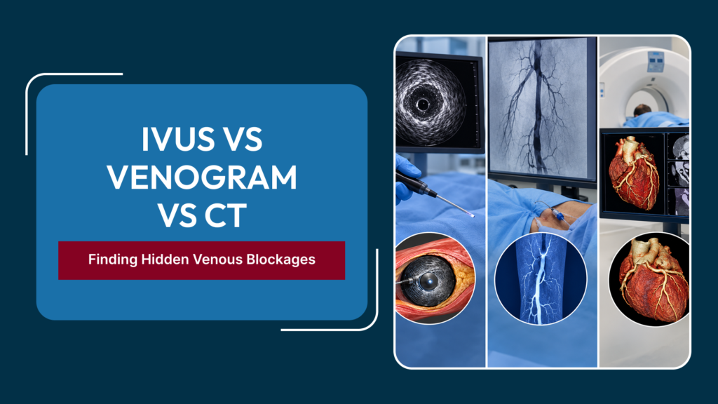
When a patient is referred for evaluation of suspected iliac vein compression, chronic venous obstruction, or recurrent DVT with unclear anatomy, the question of which imaging tests are needed — and in what sequence — is not always straightforward. Three modalities dominate the diagnostic and procedural planning process for venous blockages: CT venography, conventional venography (venogram), and intravascular ultrasound (IVUS). Each sees the venous anatomy from a different vantage point, with different strengths, limitations, and roles in the decision-making process. An Endovascular Surgeon In Delhi experienced in treating complex venous disease understands that these tests complement each other rather than substituting for one another, and that the most accurate pre-stenting assessment typically combines two or more of them rather than relying on any single modality alone.
Why Standard Duplex Ultrasound Has Limitations for Iliac Vein Assessment
Duplex Doppler ultrasound is indispensable for assessing the superficial venous system, the femoropopliteal deep veins, and venous reflux throughout the leg. It is the first-line tool for diagnosing DVT in the femoral and popliteal veins and for evaluating varicose vein incompetence. However, it has a significant anatomical blind spot: the common iliac vein and the inferior vena cava.
These structures lie deep within the pelvis, behind bowel loops that scatter ultrasound waves and obscure the vein wall. Even in ideal conditions with a cooperative patient and an experienced sonographer, the common iliac vein and the iliac vein compression point in May-Thurner Syndrome are frequently not visualised adequately. This means that the most important anatomical question — is the iliac vein compressed, narrowed, or obstructed? — cannot be reliably answered by the test that is most commonly ordered to evaluate venous disease. Cross-sectional imaging or invasive assessment is required to fill this gap.
CT Venography: The First Cross-Sectional View
What It Is and How It Works
CT venography involves injecting iodinated contrast material into a peripheral vein, then performing a CT scan timed to opacify the venous system during the scan acquisition. The result is a detailed cross-sectional and three-dimensional reconstruction of the veins from the inferior vena cava through the iliac and femoral segments to the knee level, displayed in their anatomical relationship to surrounding structures including arteries, lymph nodes, muscles, and bony landmarks.
What CT Venography Shows Well
CT venography is excellent at demonstrating the presence and level of venous obstruction, the surrounding anatomy that may be contributing to compression, the relationship between the iliac artery and the iliac vein at the May-Thurner crossing point, and the extent of chronic changes such as collateral vein formation, which develop in response to long-standing venous obstruction. It provides an overview of the entire venous anatomy from the abdomen to the knee in a single study, which is extremely useful for planning an endovascular procedure.
Limitations of CT Venography
CT venography involves ionising radiation and requires iodinated contrast, which carries a small risk of allergic reaction and nephrotoxicity in patients with impaired kidney function. More importantly, CT venography tends to overestimate the true lumen diameter of a compressed vein, because the patient is supine during the scan and venous pressure is equalised, partially opening a vessel that would be more significantly compressed in the upright position. This means that CT venography can underestimate the functional severity of iliac vein compression compared to IVUS performed at the time of intervention.
Conventional Venography (Venogram): Real-Time Flow Assessment
What It Is and How It Works
A conventional venogram is a fluoroscopy-guided procedure in which contrast is injected directly into a vein — usually through a catheter introduced via the popliteal or femoral vein — and the venous anatomy and flow are observed in real time under X-ray. The contrast column moving through the vein shows the lumen outline, identifies filling defects suggesting thrombus or stenosis, demonstrates collateral pathways, and reveals the direction and rate of venous drainage toward the heart.
What Venography Shows Well
Venography captures the dynamic aspect of venous flow that static cross-sectional imaging cannot — it shows how blood actually moves through a segment rather than just what the vessel looks like at a single point in time. It is performed in the same procedural session as the planned endovascular intervention, so it directly precedes and guides the therapeutic steps. Venography also allows pressure measurements to be taken across a stenosis or obstruction, which provide objective haemodynamic confirmation of significance when the anatomical appearance is equivocal.
Limitations of Venography
The major limitation of conventional venography is that it is a two-dimensional luminogram — it shows only the contrast-filled lumen and does not see the vein wall or the tissue outside the lumen. This is a critical limitation when assessing May-Thurner Syndrome, because the synechiae (fibrous spurs) that develop within the compressed iliac vein over time may not be visible as discrete filling defects on venography, even when they significantly reduce the functional cross-sectional area. Venography consistently underestimates the degree of iliac vein compression compared to IVUS.
Intravascular Ultrasound (IVUS): The Gold Standard for Vein Wall Assessment
What It Is and How It Works
Intravascular ultrasound involves advancing a miniaturised ultrasound probe — mounted on the tip of a catheter — directly inside the vein under fluoroscopic guidance. The probe emits ultrasound waves in a full 360-degree sweep around its circumference and receives the returning echoes to generate a real-time, cross-sectional image of the vein wall, its lumen, and the immediately surrounding tissues from within the vessel itself. Unlike any external imaging technique, IVUS sees the vein from the inside, providing information that simply is not available from CT or standard venography. A Best Endovascular Surgeon In Delhi performing venous stenting will typically use IVUS as an integral part of the procedure rather than an optional adjunct.
What IVUS Shows That Nothing Else Can
IVUS directly measures the true cross-sectional area of the vein lumen at every point along its course. It visualises the vein wall thickness, identifies intraluminal synechiae that are invisible on venography, shows the external compression from the overlying artery in real time, and allows accurate measurement of the degree of stenosis as a percentage of the normal vein cross-section. Studies consistently show that IVUS detects clinically significant iliac vein stenosis in a substantial proportion of patients in whom venography and CT venography have appeared normal or only mildly abnormal.
How IVUS Guides Stent Placement
Before stenting, IVUS defines exactly where the narrowing begins and ends, how severe it is, and whether it extends into the inferior vena cava proximally. After balloon angioplasty and stent deployment, IVUS confirms that the stent is fully expanded against the vein wall at every point, identifies any segments of incomplete apposition, and ensures the stent extends beyond the compression point at both ends to achieve an adequate seal. This post-deployment IVUS check is associated with better long-term stent patency because poorly expanded stent segments are a common cause of early stent thrombosis.
How an Endovascular Surgeon Uses These Tests Together
In clinical practice, a thoughtful pre-procedure workup for suspected iliac vein compression or chronic venous obstruction typically follows a logical sequence. CT venography or MR venography provides the initial anatomical overview and confirms that iliac vein pathology is present, justifying proceeding to an interventional assessment. Conventional venography at the time of the procedure provides real-time dynamic flow information and allows pressure measurements to confirm haemodynamic significance. IVUS performed immediately before and after stenting provides the precise, three-dimensional wall assessment that guides stent selection, sizing, and deployment, and confirms the adequacy of the result before the catheter is removed.
This multi-modality approach may seem elaborate from the patient’s perspective, but it reflects the real complexity of iliac vein anatomy and the consequences of an incorrect stent position or size. A stent placed in the wrong location, of the wrong diameter, or not fully expanded is associated with significantly higher rates of early thrombosis and recurrent obstruction — exactly the outcomes that both patient and surgeon are trying to avoid. This is why the experience and training of the Vascular Surgeon In Delhi performing the procedure matters as much as the imaging technology available.
When Is CT Venography Sufficient Without IVUS?
CT venography alone may be sufficient in a pre-procedural planning context when the degree of obstruction is clearly severe and unambiguous on cross-sectional imaging, and when the procedural goal is straightforward recanalization rather than precise characterisation of the compression degree. However, for assessment of intermediate stenoses, confirmation of May-Thurner Syndrome severity, planning of stent length and diameter, and post-deployment confirmation, IVUS adds important information that consistently changes management decisions compared to venography or CT alone.
Frequently Asked Questions
Is IVUS painful or uncomfortable?
IVUS is performed inside the vein using a catheter that is already in place for the procedure. Patients under local anaesthesia and sedation typically report no pain from the IVUS catheter itself.
Does CT venography expose me to significant radiation?
CT venography involves a radiation dose comparable to other CT studies of the abdomen and pelvis. The exact dose depends on the scanner and protocol used. For patients of reproductive age, MR venography (which does not use ionising radiation) is a useful alternative when available.
Can I have CT venography if I have impaired kidney function?
Standard CT venography uses iodinated contrast, which can worsen kidney function in susceptible patients. MR venography using gadolinium contrast or non-contrast-enhanced MR techniques may be alternatives. This should be discussed with the imaging team and the treating surgeon before the scan is ordered.
Why can’t a regular ultrasound diagnose my iliac vein problem?
Standard peripheral duplex ultrasound does not reliably visualise the common iliac vein due to its depth and the overlying bowel gas. Cross-sectional imaging (CT or MR venography) or direct intravascular assessment (IVUS or venography) is needed to evaluate this region.
How soon after CT venography will I know if I need a stent?
CT venography results are typically reviewed at the same or a subsequent consultation, and a decision about proceeding to endovascular intervention can usually be made within one to two weeks. The stenting procedure is then scheduled, typically as a day-case or overnight admission, combining diagnostic venography and IVUS with therapeutic stenting in the same session if the anatomy confirms intervention is warranted. Recovery details are covered in our guide on life after DVT thrombectomy and venous stenting.
Conclusion
IVUS, venography, and CT venography are complementary rather than competing tools, each providing a different and essential view of venous anatomy that the others cannot fully replace. An experienced Best Endovascular Surgeon In Delhi uses them in a deliberate sequence that maximises diagnostic accuracy, ensures correct stent sizing and placement, and minimises the risk of procedural error or early stent failure. Understanding why each test is used, what it shows, and what its limitations are helps patients engage meaningfully in their own diagnostic and treatment journey rather than experiencing these tests as an opaque sequence of procedures with unexplained purpose.
Find Out If Hidden Venous Blockages Are Causing Your Symptoms
If you have unexplained leg swelling, recurrent DVT, or varicose veins returning after treatment, request an appointment with our endovascular team for a structured diagnostic assessment and clear guidance on whether intervention is appropriate for your specific venous anatomy.