
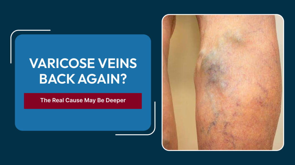
Varicose vein treatments — including endovenous laser ablation, radiofrequency ablation, foam sclerotherapy, and surgical stripping — are effective, well-validated procedures for the vast majority of patients. Most people who undergo treatment experience a sustained, durable result with good cosmetic and symptomatic improvement. Yet a meaningful subset of patients finds that varicose veins return within one to three years of treatment, sometimes more extensively than before. For many of these patients, the recurrence is attributed to the development of new tributaries or inadequate treatment of existing tributaries, and a repeat session of the same treatment is recommended. This explanation is often correct — but not always. A significant number of recurrent varicose vein cases involve an underlying deep venous or iliac vein problem that, if not identified and corrected, guarantees that superficial vein treatment will continue to fail regardless of how many times it is repeated. Knowing when a Varicose Veins Doctor In Delhi should look beyond the superficial veins is the key to breaking this cycle.
The Plumbing Principle: Why Deep Vein Problems Drive Superficial Vein Failure
To understand why deep vein disease causes superficial varicose veins, it helps to think of the venous system in terms of pressure and drainage. The deep veins are the major drainage system — responsible for carrying the bulk of the blood returning from the legs toward the heart. The superficial veins (including the great saphenous and small saphenous systems) connect to the deep system through perforator veins and join the deep veins at junction points in the groin and behind the knee.
When the deep venous system is working well, the pressure in it falls with exercise as the calf muscle pump drives blood upward. When deep venous pressure is chronically elevated — because of venous obstruction, valve incompetence from prior DVT, or extrinsic compression — that pressure is transmitted backward through the perforator veins into the superficial system. The superficial veins dilate under the increased pressure load, causing the same varicose vein pattern seen in primary disease but driven by a completely different mechanism. Treating these secondary varicosities by ablating or removing the superficial vein does temporarily remove the dilated vessel, but the source of the elevated pressure — the dysfunctional deep system — is unchanged and quickly recruits the next available superficial vein pathway.
The Three Deep Venous Problems Most Commonly Missed Behind Recurrent Varicose Veins
1. Post-Thrombotic Deep Venous Insufficiency
Prior DVT, even DVT that was treated and apparently resolved, often leaves lasting valve damage in the deep veins that causes chronic venous hypertension — the core mechanism behind post-thrombotic syndrome. Patients who had a DVT years or even decades earlier, particularly in the femoral or popliteal vein, may develop varicose veins or recurrent varicose veins as a late manifestation of this deep venous insufficiency. The history of prior DVT is often not volunteered during a varicose vein consultation, either because the patient has forgotten, because the DVT was mild and went untreated, or because the connection between the two is not recognised.
2. Iliac Vein Compression (May-Thurner Syndrome)
Compression of the left common iliac vein between the right common iliac artery and the lumbar spine — May-Thurner Syndrome — creates a chronic outflow obstruction that elevates pressure throughout the entire left leg’s venous system. Patients with this condition frequently develop left-sided varicose veins, left leg swelling, and left-sided DVT. When the varicose veins are treated without first identifying and relieving the iliac vein compression, the elevated pressure rapidly drives recurrence in adjacent superficial pathways.
3. Non-Thrombotic Iliac Vein Lesions
Not all iliac vein narrowings are caused by acute DVT or by the specific anatomy of May-Thurner compression. Some patients have primary vein wall abnormalities, extrinsic pelvic masses, or adhesive disease from prior surgery or inflammation that narrows the iliac vein without a history of acute thrombosis. These non-thrombotic iliac vein lesions (NIVILs) produce the same haemodynamic consequence — chronic venous hypertension in the ipsilateral leg — and the same pattern of recurrent varicose vein failure.
Clinical Clues That Should Trigger Deep Vein Investigation
- Varicose veins recurring within two years of technically successful ablation or surgery.
- Varicose veins predominantly or exclusively affecting the left leg.
- Varicose veins with associated persistent leg swelling or skin changes disproportionate to the size of the visible varicosities.
- A history of DVT at any point in the past, even if considered remote and fully resolved.
- Varicose veins that developed or worsened significantly during or after pregnancy.
- Varicose veins with an atypical distribution — originating from the pelvis rather than the groin junction, or presenting along the inner thigh with no clear saphenous junction reflux on duplex.
- Pelvic varicose veins visible in the distribution of ovarian or internal iliac tributaries, suggesting pelvic venous insufficiency as a driver.
What Investigations Are Needed?
A standard duplex Doppler ultrasound performed by a skilled vascular sonographer is the first step, and it should extend beyond the groin junction to assess deep vein reflux and, where possible, the iliac vein. However, the iliac vein at its proximal extent is beyond the reliable reach of standard peripheral ultrasound, and additional cross-sectional imaging is usually needed when the clinical suspicion for deep venous obstruction is high. CT venography, MR venography, and — at the time of intervention — intravascular ultrasound are the modalities used to precisely characterise iliac vein pathology. Our comprehensive guide on IVUS versus venogram versus CT venography for diagnosing venous blockages covers each of these in detail, explaining what each test offers and how they are used together to plan treatment.
Treatment When Deep Vein Disease Is Confirmed
The treatment approach depends entirely on the mechanism identified. If the deep venous problem is primarily reflux from damaged valves after prior DVT, compression therapy forms the foundation of management alongside any repeated superficial treatment, since there is currently no durable valve repair technique in routine clinical use. If the deep venous problem is obstruction — iliac vein compression, residual organised thrombus, or another outflow block — endovascular stenting to restore the outflow pathway is the definitive treatment, and it should precede or accompany any further superficial vein procedure.
Proceeding with repeat superficial ablation before relieving confirmed outflow obstruction is a sequencing error that predictably leads to early recurrence and patient frustration. The correct sequence is to diagnose and treat the proximal obstruction first, allow the venous haemodynamics to normalise over weeks to months, and then reassess whether superficial treatment is still needed and what exactly remains to treat. Some patients find that their superficial varicosities partially involute once the outflow obstruction is relieved, reducing the extent of additional superficial treatment required. What the post-stenting monitoring and anticoagulation protocol looks like is described in our article on life after DVT thrombectomy and venous stenting.
Frequently Asked Questions
My varicose veins came back a year after laser treatment — does that always mean a deep vein problem?
Not always. Early recurrence is sometimes due to incomplete treatment of the original saphenous reflux or rapid development of tributaries. However, if recurrence is on the left side, involves unusual vein distributions, or is accompanied by swelling, a deep vein evaluation is worthwhile before repeating superficial treatment.
Can an ultrasound performed at my varicose vein clinic detect iliac vein problems?
Standard varicose vein clinic duplex scanning focuses on the superficial and perforator systems and often does not include a detailed assessment of the iliac veins. Dedicated imaging — either a specialist-requested duplex or CT/MR venography — is required when iliac vein obstruction is suspected.
Is iliac vein stenting a major operation?
No. Like most modern endovascular procedures, iliac vein stenting is performed through a small catheter access site, typically under local anaesthesia and sedation, without any open surgical incision. Recovery is substantially faster than open vascular surgery. Details of the procedure and recovery are covered in our post-procedure guide on life after DVT thrombectomy and venous stenting.
I had a DVT ten years ago and now have varicose veins for the first time — could they be related?
Yes. Post-thrombotic deep venous insufficiency can manifest years after the original DVT, and varicose veins developing in this context are secondary to the chronic deep venous pressure elevation. This is precisely the kind of history that should prompt a thorough deep vein assessment before any superficial varicose vein treatment. Read more about post-thrombotic syndrome for a detailed explanation of how prior DVT causes lasting venous problems.
Will treating my iliac vein problem cure my varicose veins permanently?
Relieving a confirmed iliac vein obstruction corrects the source of elevated venous pressure that was driving varicose vein recurrence. This does not guarantee that all existing superficial varicosities will disappear, but it creates the haemodynamic conditions in which subsequent superficial treatment is much more likely to be durable.
Conclusion
Varicose vein recurrence after treatment is not inevitable, but when it does happen — particularly in predictable patterns involving the left leg, atypical distribution, disproportionate swelling, or a history of DVT — it is a signal that demands a thorough deep vein and iliac vein assessment rather than an immediate repeat of the same superficial treatment. A Varicose Veins Doctor In Delhi who understands both the superficial and deep venous systems, and who has access to advanced imaging and endovascular correction of deep venous pathology, offers patients with recurrent disease the most accurate diagnosis and the most durable long-term outcome.
Varicose Veins Returning? Get a Complete Venous Assessment
Do not repeat superficial treatment without ruling out deep vein disease. Book a comprehensive venous evaluation with our specialist team for an accurate diagnosis and a treatment plan designed to address the root cause of your recurrence.