For patients on long-term hemodialysis, the quality of their vascular access has a direct bearing on how effective and comfortable each dialysis session is, which is why access preservation is treated as seriously as the dialysis prescription itself. An arteriovenous (AV) fistula — a surgical connection between an artery and a vein, usually in the arm — is considered the gold-standard access for hemodialysis because it offers durable, high-flow blood access with fewer complications than a catheter or graft. But fistulas are not maintenance-free: over months to years, narrowing (stenosis) can develop at the site of the surgical connection or along the outflow vein, threatening the ability to dialyze effectively. AV fistula angioplasty in Delhi is the primary tool used to reopen a narrowed fistula and preserve this lifeline access without needing to create a new one.
Why Fistulas Develop Stenosis
The most common site of narrowing is the venous anastomosis — the point where the vein was surgically joined to the artery — where turbulent, high-pressure arterial flow triggers a thickening of the vessel wall known as intimal hyperplasia. Stenosis can also develop further along the outflow vein, sometimes at old needle-puncture sites where repeated cannulation has caused scarring, or centrally in the chest veins if the patient has had previous catheters. Over time this narrowing reduces flow through the fistula, which can compromise dialysis efficiency long before the fistula fails outright.
Signs a Fistula Needs Attention
- A weaker or absent thrill (the buzzing vibration normally felt over a working fistula)
- Prolonged bleeding from needle sites after dialysis
- Rising venous pressures recorded during dialysis sessions
- Falling access flow rates on routine surveillance
- New swelling in the arm, hand, or shoulder
- Difficulty achieving the prescribed dialysis dose despite adequate needle placement
How Stenosis Is Diagnosed
Physical examination — feeling for the thrill and listening for the bruit (the whooshing sound of flow) along the fistula — remains a quick bedside screen for a problem. Duplex ultrasound is used to measure flow volume and locate areas of narrowing non-invasively, and when intervention is being planned, a fistulogram (venography performed by injecting contrast dye into the access) gives a precise picture of the stenosis before treatment.
When Angioplasty Is Actually Required
Not every narrowing seen on imaging needs to be treated. Intervention is generally reserved for a stenosis that is both anatomically significant — typically reducing the vessel diameter by more than half — and clinically significant, meaning it is accompanied by reduced access flow, difficult cannulation, rising dialysis venous pressures, or arm swelling. Treating an incidental narrowing that is not causing any functional problem is usually avoided, since repeated interventions can themselves contribute to further scarring.
The Angioplasty Procedure
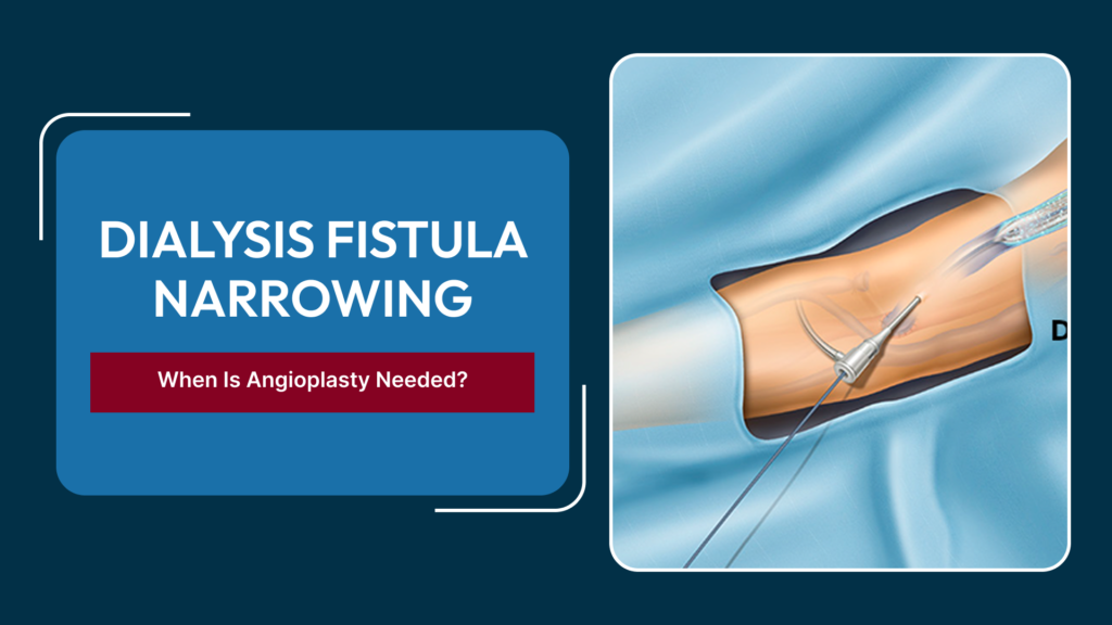
Fistula angioplasty is done percutaneously — a needle and guidewire access the fistula, a balloon catheter is advanced to the narrowed segment under imaging guidance, and the balloon is inflated to stretch the vessel back open. For stenosis that recurs quickly after plain balloon angioplasty, a drug-coated balloon may be used to reduce the rate of renarrowing. If the segment keeps collapsing back (elastic recoil) or the narrowing is in a central vein where balloon angioplasty alone is insufficient, a stent may be placed to hold the vessel open. Most procedures are done under local anesthesia and patients typically go home the same day.
When Angioplasty Is Not Enough
Some fistulas develop stenosis repeatedly at the same site despite angioplasty, or the vein becomes too scarred to treat effectively with a balloon. In these situations, surgical revision — moving the connection to a fresh segment of vein, or creating a new fistula or graft altogether — may be recommended. The goal throughout is to preserve as much of the patient’s own vein anatomy as possible, since access sites are a limited resource over a dialysis patient’s lifetime.
A Related but Distinct Condition: AV Malformations
It’s worth distinguishing a dialysis AV fistula from an arteriovenous malformation (AVM), which is a separate, usually congenital, abnormal tangle of arteries and veins that has not been surgically created. AVMs are managed differently, often with embolization, sclerotherapy, or surgical excision rather than angioplasty. Patients seeking AV malformation treatment in Delhi for a congenital vascular lesion should have it evaluated separately from any dialysis access issue, since the underlying anatomy and treatment goals are not the same.
Why Surveillance Matters More Than One-Time Treatment
A single successful angioplasty does not make a fistula maintenance-free. Because the same forces that caused the original stenosis — turbulent flow and repeated needle trauma — continue after treatment, ongoing surveillance through monthly physical examination and periodic flow measurement is what catches recurrence early, while it can still be treated with a straightforward balloon angioplasty rather than an emergency intervention on a fistula that has already clotted off. Access centers that build this surveillance into routine dialysis care tend to see fewer catheter placements and fewer lost accesses over time.
What Happens If a Fistula Clots Completely
If a stenosis is left untreated long enough, the fistula can clot off entirely, at which point angioplasty alone is no longer sufficient. Thrombectomy — mechanically or pharmacologically clearing the clot — combined with angioplasty of the underlying stenosis can sometimes salvage even a fully clotted access if performed promptly, but success rates fall the longer the fistula has been non-functional. This is another reason why symptoms of a failing fistula are treated as a priority rather than something to monitor over several dialysis sessions.
Frequently Asked Questions
How often should a dialysis fistula be checked for narrowing?
Most dialysis units perform routine physical examination at every session and periodic flow surveillance, with further imaging triggered whenever the thrill weakens, dialysis pressures rise, or flow rates fall.
Does fistula angioplasty require a hospital stay?
No, it is usually done as a day procedure under local anesthesia, and most patients can resume dialysis through the same access shortly afterward.
How long does the benefit of angioplasty last?
It varies by patient and location of the stenosis; some fistulas stay open for a year or more after a single angioplasty, while others narrow again within months and need repeat treatment or a drug-coated balloon.
Can a fistula be saved if angioplasty stops working?
Often yes — options include repeat angioplasty, a drug-coated balloon, stenting, or surgical revision of the access before resorting to creating an entirely new fistula or graft.
Is an AV malformation the same as a dialysis fistula?
No — an AV malformation is a separate, typically congenital, abnormal connection between arteries and veins, while a dialysis fistula is deliberately created by surgery for dialysis access; the two are evaluated and treated differently.
Protecting a working dialysis fistula starts with catching narrowing early. For evaluation of access dysfunction or a suspected vascular malformation, visit our vascular surgery FAQs