The standard first response to a deep vein thrombosis diagnosis is anticoagulation — blood thinners administered either as an injection or a tablet, with the goal of preventing the existing clot from growing while the body gradually dissolves it on its own. For the majority of patients with small or distal DVT confined to the calf veins, this approach works well and carries an acceptable risk profile. But for a significant and often underappreciated subset of patients, blood thinners alone are an incomplete answer — one that prevents death from pulmonary embolism while leaving behind a damaged, partially obstructed venous system that causes years of chronic suffering. This is where clot suction for DVT and mechanical thrombectomy have fundamentally changed what is possible in DVT care.
What Blood Thinners Do — and What They Cannot Do
Anticoagulants work by interrupting the clotting cascade, preventing new thrombus from forming and allowing the body’s natural fibrinolytic system to gradually break down the existing clot. The critical limitation is that this natural dissolution process is slow, unpredictable, and often incomplete. Studies consistently show that a significant proportion of patients treated with anticoagulation alone retain residual thrombus — permanent scarring and obstruction within the deep vein — even after months of treatment. This residual damage triggers the chronic venous hypertension that underlies post-thrombotic syndrome.
Post-thrombotic syndrome affects an estimated thirty to fifty percent of DVT patients managed with anticoagulation alone, producing years of leg heaviness, swelling, pain, skin changes, and in severe cases, chronic venous ulcers that are difficult and expensive to manage. The fundamental problem is that blood thinners stop the clotting process but do not actively remove the clot that has already formed. Understanding this distinction is the starting point for understanding when a more active, mechanical approach — performed by a Best DVT Treatment Doctor In Delhi with endovascular expertise — becomes the better clinical choice.
What Is Clot Suction for DVT?
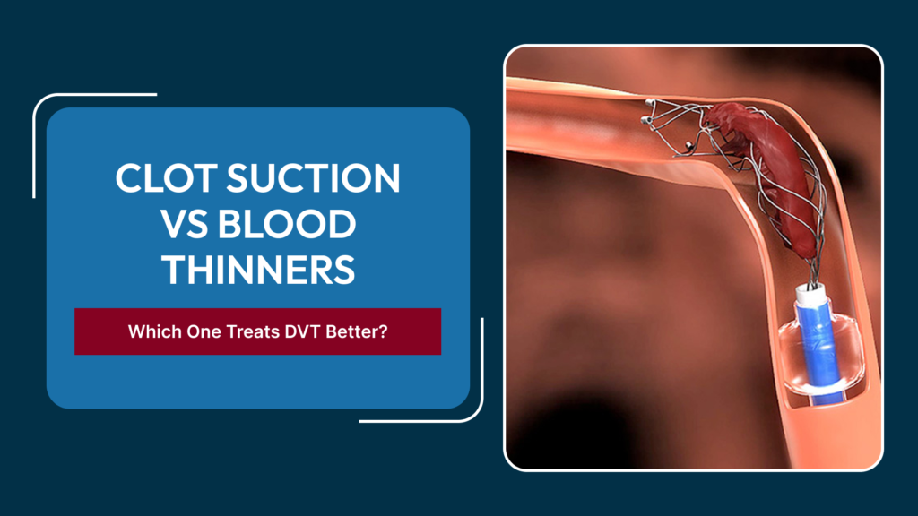
Clot suction — more formally called pharmacomechanical catheter-directed thrombectomy or simply percutaneous mechanical thrombectomy — is a minimally invasive endovascular procedure in which a catheter is introduced into the thrombosed vein, and the clot is either broken up mechanically, aspirated under suction, dissolved using locally delivered thrombolytic drugs, or treated with a combination of all three approaches. The goal is direct, rapid removal of the thrombus from the affected vein segment rather than waiting for the body’s own dissolution system to work gradually and incompletely over weeks or months.
Modern thrombectomy systems are designed to minimise bleeding risk by localising the thrombolytic drug delivery directly to the clot rather than infusing it systemically. Some of the newest generation devices use only mechanical suction without any pharmacological thrombolysis at all, further reducing the bleeding risk that was the historic barrier to offering this approach more broadly.
Who Is a Candidate for Mechanical Thrombectomy Rather Than Anticoagulation Alone?
Ilio-Femoral DVT in Young, Active Patients
The strongest evidence for early clot removal applies to patients with ilio-femoral DVT — clot extending from the thigh into the iliac vein — who are young and otherwise healthy enough to tolerate the procedure. These patients stand to lose the most from incomplete venous clearance, since they have decades ahead of them in which post-thrombotic syndrome would significantly impair quality of life. A Best Vascular Surgeon In Delhi experienced in endovascular techniques will typically consider thrombectomy strongly in this group, particularly when symptoms are severe and the clot is recent — ideally less than fourteen days old.
Acute Phlegmasia Cerulea Dolens
This is the most urgent presentation of DVT — massive ilio-femoral thrombosis causing such extreme venous outflow obstruction that limb viability is threatened. The leg becomes intensely swollen, cyanotic (blue-purple), and acutely painful, with arterial flow ultimately compromised by compartment pressure. This is a vascular emergency where mechanical thrombectomy is not merely preferred but urgently required, often combined with fasciotomy, to save the limb.
Symptomatic DVT With Underlying Structural Cause
When DVT occurs on the background of a structural venous problem — most commonly May-Thurner Syndrome with iliac vein compression — removing the acute clot is only half the treatment. The residual compressed segment must then be stented to maintain patency. In these cases, thrombectomy is performed specifically to clear the vein of acute thrombus so that accurate assessment of the underlying anatomy is possible and a stent can be placed correctly without clot interfering with the result.
Failed or Incomplete Response to Anticoagulation
Patients who present with persistent or worsening symptoms despite adequate anticoagulation, or who have significant residual thrombus burden on repeat imaging several weeks into treatment, may be candidates for delayed thrombectomy to remove the remaining organised clot before it becomes fully fibrosed and permanently incorporated into the vein wall.
Patients Who Cannot Tolerate Long-Term Anticoagulation
Paradoxically, patients with high bleeding risk who are poor candidates for prolonged anticoagulation may sometimes benefit from early mechanical thrombectomy, which provides definitive clot removal more rapidly and may allow a shorter overall anticoagulation duration compared to the watch-and-wait approach.
The Procedure: What Happens During Clot Suction for DVT?
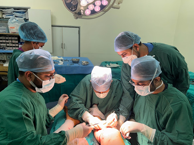
The procedure is performed in an endovascular suite under fluoroscopic guidance, typically with the patient under local anaesthesia and conscious sedation or light general anaesthesia. Access is usually obtained through the popliteal vein behind the knee or through a femoral access site, depending on the extent and location of the clot. A venogram is performed first to map the extent of thrombosis and identify any underlying anatomical abnormality.
The thrombectomy catheter is then advanced through the clot under imaging guidance. Depending on the device used, the catheter either mechanically macerates and aspirates the thrombus, delivers a lytic agent directly into the clot while simultaneously fragmenting it, or uses ultrasound-assisted technology to enhance drug penetration into the clot. Repeat venography is performed during the procedure to assess clearance, and a venous stent is placed at the same sitting if significant residual stenosis or extrinsic compression is identified.
A detailed description of the imaging and diagnostic steps that precede stent placement — including how IVUS, venography, and CT venography are used together — is covered in our specialist guide on how an endovascular surgeon in Delhi diagnoses hidden venous blockages before stenting.
Recovery and What to Expect Afterwards
Most patients undergoing catheter-directed thrombectomy stay in hospital overnight or for one to two nights for monitoring and continuation of anticoagulation. The leg typically shows measurable improvement in swelling within the first twenty-four to forty-eight hours after successful clot removal, which is a sharp contrast to the gradual, incomplete resolution seen with anticoagulation alone. Anticoagulation continues after the procedure for a defined period, and follow-up imaging at specified intervals confirms ongoing patency. The complete post-procedure roadmap — including travel restrictions, compression garment use, and when follow-up Doppler or venography is scheduled — is described in our article on life after DVT thrombectomy and venous stenting.
Comparing Outcomes: Clot Suction Versus Anticoagulation Alone
- Clot removal: Mechanical thrombectomy removes the bulk of the clot during the procedure; anticoagulation relies on gradual, incomplete natural dissolution over weeks.
- Post-thrombotic syndrome: Early thrombectomy significantly reduces the risk and severity of post-thrombotic syndrome compared to anticoagulation alone in ilio-femoral DVT.
- Symptom relief: Swelling and pain improve faster after thrombectomy, typically within days rather than weeks.
- Venous patency: Veins treated with early thrombectomy and stenting where indicated show substantially better long-term patency rates than veins managed conservatively.
- Bleeding risk: Modern pharmacomechanical systems carry lower systemic bleeding risk than older catheter-directed thrombolysis infused over many hours, though the procedure is not risk-free and patient selection matters enormously.
Frequently Asked Questions
Is clot suction for DVT a painful procedure?
The procedure is performed under local anaesthesia and conscious sedation or general anaesthesia, so there is no significant pain during the procedure. Post-procedurally, the leg may feel sore and bruised, but most patients report significant relief of the pre-procedure heaviness and swelling within one to two days.
How soon after DVT diagnosis should thrombectomy be performed?
Earlier is better. Acute clot — typically less than fourteen days old — responds best to mechanical removal. Older, organised clot is firmer and less amenable to suction or fragmentation, though even partially organised thrombus can sometimes be addressed with appropriate techniques.
Can clot suction for DVT be performed if I am already on blood thinners?
Yes. Anticoagulation is typically started immediately upon DVT diagnosis, and the endovascular team will manage the anticoagulation around the procedure window. Being on blood thinners does not disqualify a patient from thrombectomy.
Are there patients for whom mechanical thrombectomy is not suitable?
Patients with active bleeding, recent stroke, uncontrolled severe hypertension, or certain other conditions may not be candidates. Patient selection is based on a careful risk-benefit assessment by the treating endovascular team.
Does successful clot removal mean I will not get DVT again?
Not necessarily. If the DVT occurred on the background of an underlying structural problem like May-Thurner Syndrome, treating that structural cause through stenting is essential to prevent recurrence. Anticoagulation may also continue long-term depending on individual thrombotic risk factors.
Conclusion
Blood thinners remain an essential part of DVT management, but they are not always sufficient on their own — particularly for extensive ilio-femoral DVT, where the consequences of incomplete venous clearance can mean decades of chronic post-thrombotic suffering. Mechanical thrombectomy and clot suction represent a meaningful advance that gives patients with the right clinical profile the opportunity for more complete venous restoration and a significantly lower risk of long-term disability. The decision requires the expertise of a specialist in vascular and endovascular surgery who can assess the full clinical picture rather than defaulting to the same anticoagulation protocol applied to all presentations of DVT.
Speak to a DVT Treatment Specialist in Delhi
If you have extensive DVT, recurrent DVT, or symptoms that have not improved adequately on blood thinners, request a consultation to discuss whether clot suction or endovascular intervention is appropriate for your specific situation.