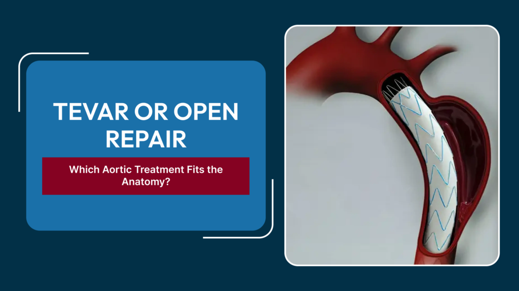
Thoracic aortic aneurysms and dissections can be treated with two fundamentally different approaches: thoracic endovascular aortic repair (TEVAR), which places a stent graft from within the vessel through a small groin incision, or open surgical repair, which replaces the diseased segment of aorta directly through the chest. Both aim to prevent rupture or further dissection, but they are not interchangeable — the aorta’s anatomy in each patient often makes one approach clearly preferable, or the only viable option. An endovascular surgeon in Delhi weighs a detailed set of anatomical measurements before recommending either route.
What TEVAR Involves
In TEVAR, a fabric-covered stent graft is folded into a delivery catheter and advanced from an artery in the groin up to the diseased segment of the thoracic aorta, where it is deployed to line the vessel wall and exclude the aneurysm or dissection from blood pressure. Because there is no chest incision and no need for cardiopulmonary bypass in most cases, TEVAR is generally associated with a shorter hospital stay, less blood loss, and a faster return to normal activity than open surgery. It has become the preferred first option whenever the anatomy allows it.
What Open Repair Involves
Open repair involves a thoracotomy (an incision into the chest) to directly access the aorta, clamp it above and below the diseased segment, and replace that segment with a synthetic graft sewn in place. Depending on the extent of disease and its proximity to the heart, this may require cardiopulmonary bypass and periods of circulatory arrest. Recovery is longer and the perioperative risk profile is higher than TEVAR, but open repair remains definitive for anatomy that a stent graft cannot safely treat, and it avoids some of the long-term surveillance concerns that come with an endovascular repair.
Anatomical Factors That Decide the Approach
Landing Zone Length and Quality
TEVAR requires a healthy, adequately long segment of aorta above and below the diseased area — the ‘landing zones’ — to seat the stent graft securely and create a durable seal. Short, angulated, or heavily calcified landing zones increase the risk of the graft not sealing properly, sometimes ruling out an endovascular approach or requiring more complex grafts.
Involvement of Arch Branches
When an aneurysm or dissection extends into the aortic arch and involves the branches supplying the brain and arms, a standard stent graft would block these vessels. This anatomy either requires specialized branched or fenestrated stent grafts, a hybrid procedure combining surgical debranching with endovascular repair, or open arch replacement, depending on what the specific branch pattern allows.
Access Vessel Size and Tortuosity
TEVAR devices are delivered through the femoral or iliac arteries, so these vessels need to be large enough and not too tortuous or calcified to pass the delivery system safely. Small or severely diseased access vessels can make a purely percutaneous approach difficult, sometimes requiring a surgical conduit to gain access.
Connective Tissue Disorders
Patients with Marfan syndrome, Ehlers-Danlos syndrome, or other connective tissue disorders tend to have progressive, diffuse aortic dilation rather than a localized aneurysm, and stent grafts anchored in fragile, still-dilating aorta have a higher long-term failure rate in this group. Open repair is generally favored in confirmed connective tissue disease when the patient is fit for surgery.
Aneurysm vs Dissection
Acute dissections, especially those involving the ascending aorta, are usually managed with emergency open surgery, while dissections confined to the descending aorta with complications such as malperfusion or impending rupture are often well suited to TEVAR. Chronic, stable dissections and degenerative aneurysms are assessed on their own anatomical merits.
Patient Factors Beyond Anatomy
Age, cardiac and pulmonary function, prior chest or abdominal surgery, and overall fitness for a major operation all factor into the decision alongside anatomy. A patient with anatomy suited to either approach but significant comorbidities may still be steered toward TEVAR specifically to reduce perioperative risk, even when open repair would otherwise be a reasonable option.
Follow-Up Differs Between the Two
TEVAR patients need lifelong periodic imaging, typically CT scans, to check for endoleak — persistent blood flow into the aneurysm sac around the graft — and to confirm the graft has not migrated. Open repair patients are also followed long-term, but the surveillance burden and specific complications monitored for differ, since the failure modes of a sewn graft are not the same as those of a stent graft.
Why Planning Imaging Takes Time
Because the margin for error in aortic measurements is small — a landing zone that is a few millimetres too short can mean the difference between a durable seal and a late endoleak — planning for either TEVAR or open repair typically involves high-resolution CT angiography reviewed with dedicated measurement software, sometimes alongside 3D reconstruction to assess branch anatomy and angulation. This planning stage is not a formality; it is what determines whether an off-the-shelf stent graft will fit, whether a custom or fenestrated device needs to be ordered, or whether open repair is the only anatomically sound option, and it can take longer for complex arch or thoracoabdominal disease than for a straightforward descending aneurysm.
Emergency Presentations Change the Calculus
When a patient presents with a ruptured or rapidly expanding aneurysm, or a complicated acute dissection with malperfusion, there is little time for the extensive imaging work-up that elective planning allows. In these emergencies, the anatomy is assessed as quickly as possible and the approach most likely to control the problem fastest — often TEVAR where the anatomy permits, given its speed of deployment — is prioritized over the more deliberate, definitive planning used for elective aneurysm repair.
Frequently Asked Questions
Is TEVAR always the safer option?
TEVAR generally carries lower short-term procedural risk, but it is only safe and durable when the anatomy provides adequate landing zones and access; in unsuitable anatomy, forcing an endovascular approach can be riskier than a well-planned open repair.
How is the right approach determined before treatment?
CT angiography with detailed measurements of the aorta’s diameter, length, angulation, and branch involvement is used to map the anatomy precisely, and this imaging is what ultimately decides which approach is feasible.
Can a patient switch from TEVAR to open repair later if needed?
Yes, a stent graft that develops a significant endoleak, migrates, or fails to control the aneurysm can be treated with a further endovascular procedure or, in some cases, converted to open repair.
Does TEVAR eliminate the risk of rupture completely?
It substantially reduces the risk when the graft is well-sealed, but ongoing surveillance is needed because a minority of patients develop an endoleak or graft migration that can allow pressure to return to the aneurysm sac.
Why do some patients need a hybrid procedure?
When the diseased segment involves branches supplying the brain or arms, a hybrid approach — surgically rerouting those branches followed by stent graft placement — can extend endovascular treatment to anatomy that a standard stent graft alone could not cover.
Aortic aneurysm treatment decisions depend heavily on precise imaging and anatomy. To discuss evaluation and treatment planning, visit our vascular surgery gallery and case outcomes